The human body has 33 vertebrae and the spine is comprised of 24 of them. In the measurement of vertebral body heights the accuracy decreased at vertebral levels where the images of thoracic vertebral bodies are superimposed upon by the shadows of cardiovascular organs.

Hounsfield Units On Lumbar Computed Tomography For Predicting
Measurement of vertebral body. However differences in the range of vertebral body collapse may result in measurement errors. Coefficients of variation for repeat qm and mxa scan analysis were less than 2. The size of the vertebral bodies increases down the spine as the size and weight of the body it has to support above it increase. The five lumbar vertebral bodies found in the lower back are bigger and thicker than those located in the cervical and thoracic regions. Because the variations in measurement values in japanese were not significant in comparison with those in european and american persons means minus 3 sd were almost the same as means minus 25 designated in japan with respect to the ratio of anterior to posterior margin heights of vertebral bodies. In the human vertebral column the size of the vertebrae varies according to placement in the vertebral column spinal loading posture and pathology.
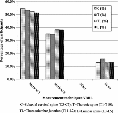
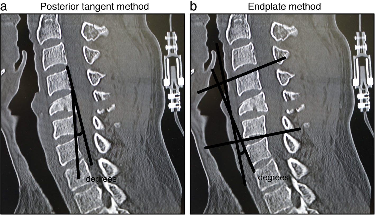
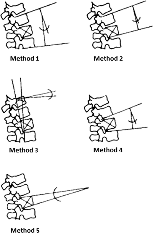
Previously researchers used different measurement methods to assess the degree of vertebral body collapse by using posterior wall height as the reference vertebral body compression ratio vbcr or the percentage of anterior height compression pahc which uses the mean height of segments adjacent to a healthy vertebral body as the reference value 811. The vertebral body is the large anterior cylindrical portion that is predominantly responsible for bearing the weight of the spine and body above it. Along the length of the spine the vertebrae change to accommodate different needs related to stress and mobility. This increased size is necessary to accommodate the weight load and pressure exerted against the lower back. Anterior middle and posterior vertebral body heights of all segments from t4 to l4 were measured interactively using mxa software and qm from the spinal radiographs and compared with direct measurements derived using digital callipers following cadaveric dissection.

