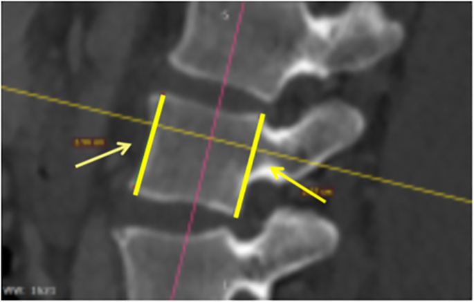
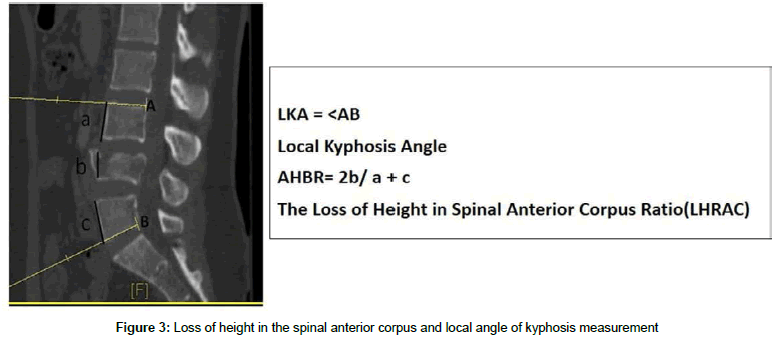
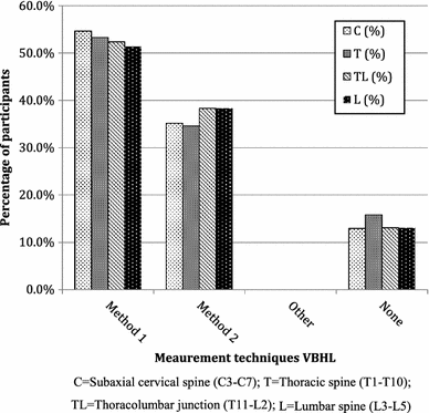
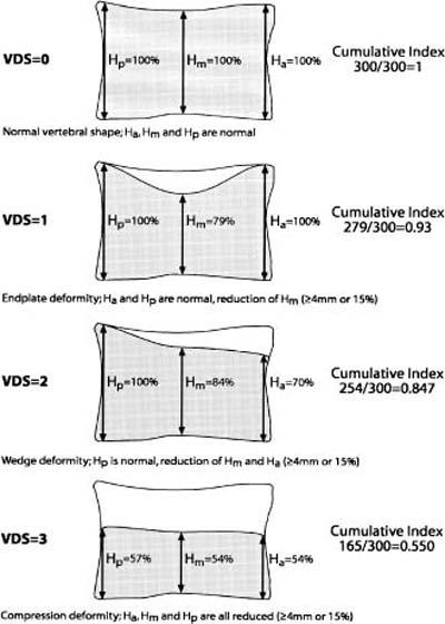
Previously researchers used different measurement methods to assess the degree of vertebral body collapse by using posterior wall height as the reference vertebral body compression ratio vbcr or the percentage of anterior height compression pahc which uses the mean height of segments adjacent to a healthy vertebral body as the reference value 8 11. Under the optimal ex vivo scanning conditions used in this study mxa is comparable to spinal radiography for the assessment of vertebral height.

View Image
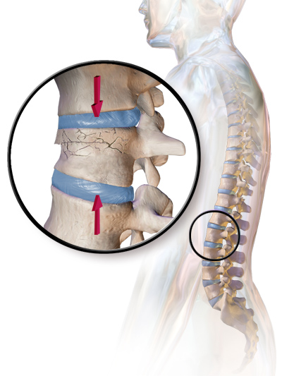
Vertebral body height measurement. A spinal cord injury sci can occur anywhere along the spinal cord and causes a loss of communication between the brain and the parts of the body below the. There was strong linear association between the mxa and qm measurements r2 099 with mean differences at the three measurement sites ranging from 41 to 59. The spinal cord itself is about 45 cm 18 in in men and 43 cm 17 in long in women. Criteria for deformity should allow for this variation to avoid misdiagnosis. Previously researchers used different measurement methods to assess the degree of vertebral body collapse by using posterior wall height as the reference vertebral body compression ratio vbcr or the percentage of anterior height compression pahc which uses the mean height of segments adjacent to a healthy vertebral body as the reference value 8. The average dorsal t12v body height was 2125164 mm in the control group and 2011149 mm in the lvf group.
The normal lower bound for relative change in measured vertebral heights ranged between 10 and 16 anteriorly and 11 and 19 posteriorly. The human spine is composed of 33 vertebrae that interlock with each other to form the spinal column. The average ventral t12v body heights were 1951154 mm and 1762195 mm respectively. The lvf group had significantly lower dorsal and ventral t12v body heights both p0001. Normal vertebral dimensions were found to vary from vertebra to vertebra.

